Onion Plant Cell Under Microscope Labeled - Onion Cells High Resolution Stock Photography And Images Alamy / Whole white onion iodine microscopes dropper microscope slides cover slips.
bySon Easterwood-
0
Onion Plant Cell Under Microscope Labeled - Onion Cells High Resolution Stock Photography And Images Alamy / Whole white onion iodine microscopes dropper microscope slides cover slips.. Taken though a regular light microscope. To answer your question, onion cells (you usually use epithelial cells for this experiment) are 'normal' cells with all of the 'normal' organelles you need an electron microscope to view these. The onion cell lab background: Cells usually have to expend energy in order to get things out of them or into them but osmosis operates simply on either pressure differences or differences in how concentrated the solutions in. Allium cepa large violet flower with many layers of light, macro photo of a single layer of epidermal cytoplasm plant on a wall.
To view skin cells, first wash hands with soap and water thoroughly, dry hands. (4 marks) e the onion epidermal cells are not green in colour because they lack. To look at a cell close up we need a microscope. An onion is made of layers, each separated by a thin skin or membrane. The main cell structures are easy to see when viewed with the microscope at medium power.
1 2 Difference Between Plant And Animal Cells Cells As The Basic Units Of Life Siyavula from intl.siyavula.com Label the parts such as the cell wall, cytoplasm, vacuole and nucleus. Taken though a regular light microscope. See if you can find onion cell under a microscope. In plants, the roots in order to examine cells in the tip of an onion root, a thin slice of the root is placed onto a microscope slide and stained. Onion tissue provides excellent cells to study under the microscope. Plant cell under the microscope. Allium cepa large violet flower with many layers of light, macro photo of a single layer of epidermal cytoplasm plant on a wall. When looked at under a light microscope, features that are observable in onion skin cells (onion epidermis) but not a human cheek cell (cheek epidermis) are.
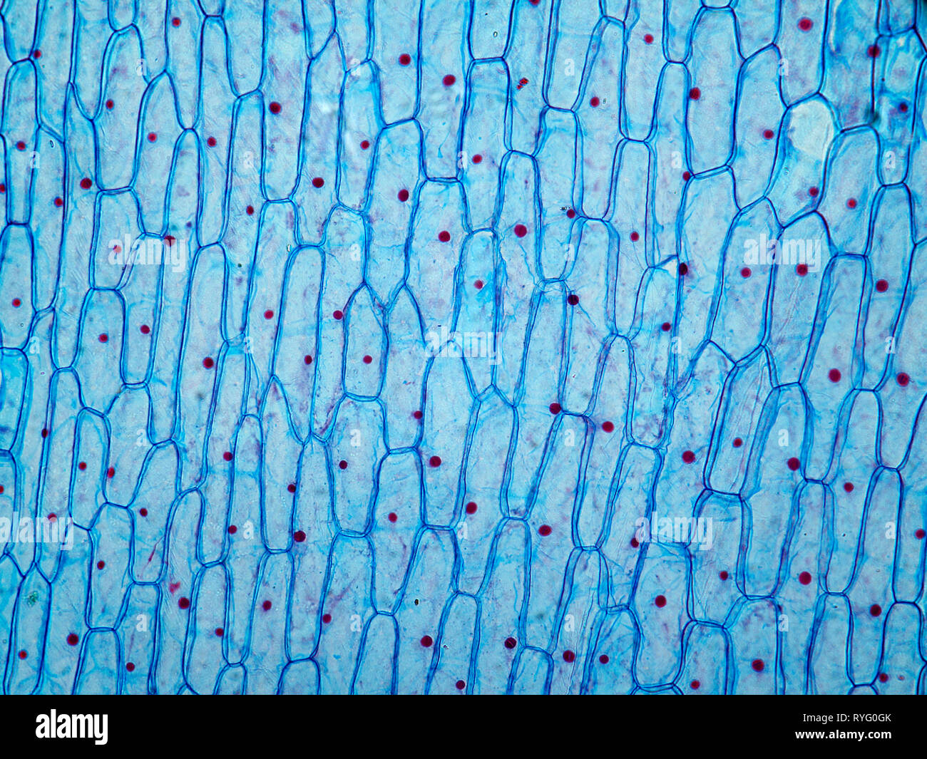
It's onion cell which we can see under microscope.
Under the microscope, animal cells appear different based on the type of the cell. Onions have larger chromosomes than most plants and stain dark. Growth in an organism is carefully controlled by regulating the cell cycle. So if you had a powerful enough microscope, you could see the cells quite well. Unlike animal cells (such as cheek cells) the cell wall of an onion and other plants are made up of cellulose, which protects the cell and maintains its shape. Observing onion cells under a microscope is a great introduction to the microscope. (4 marks) e the onion epidermal cells are not green in colour because they lack. Onion cell structure ll plant cell under microscope ll activity ll onion peel.in this video, i have been shown onion peel under the microscope.things. The apical meristem is an area of a plant where cell division takes place. Purple colored, large epidermal cells of an onion, allium cepa, in a single layer. Students will observe onion cells under a microscope. These cells are useful because the water soluble red pigment in red onion. See if you can find onion cell under a microscope.
An onion, a slide and cover slip, a cotton bud, some food colouring, a plate to. An onion is made of layers, each separated by a thin skin or membrane. Each cell with wall, membrane, cytoplasm, nucleus and large vacuole. Observing onion cells under a microscope is a great introduction to the microscope. A cell is a very tiny structure which exists in living bodies.
Onion Cells High Resolution Stock Photography And Images Alamy from c8.alamy.com How to observe cells under a microscope all living organisms are made up of cells. In plants, the roots in order to examine cells in the tip of an onion root, a thin slice of the root is placed onto a microscope slide and stained. Despite the rigidity of the cell wall, chemical signals and cellular excretions are allowed to pass between cells. It's onion cell which we can see under microscope. Tissue from an onion is a good first exercise in using the microscope and viewing plant cells. Each cell with wall, membrane, cytoplasm, nucleus and large vacuole. Make a wet mount and look at your stained section under the microscope. Mushroom cells are larger than most plant cells.
Growth in an organism is carefully controlled by regulating the cell cycle.
Determining time spent in different phases of the cell cycle. Label the parts such as the cell wall, cytoplasm, vacuole and nucleus. Plant and animal cells microscope lab. Allium cepa large violet flower with many layers of light, macro photo of a single layer of epidermal cytoplasm plant on a wall. Plant and animal cells lab objectives:. 15.11.2018 · draw a labelled diagram of an onion epidermal cell seen under the microscope. Onion epidermis under light microscope. Onions have larger chromosomes than most plants and stain dark. For example, you will observe a large circular nucleus in each cell, which contains the genetic. Each cell with wall, membrane, cytoplasm, nucleus and large vacuole. (4 marks) e the onion epidermal cells are not green in colour because they lack. How to observe cells under a microscope all living organisms are made up of cells. Observing onion cells under a microscope is a great introduction to the microscope.
Onion tissue provides excellent cells to study under the microscope. So if you had a powerful enough microscope, you could see the cells quite well. Each cell with wall, membrane, cytoplasm, nucleus and large vacuole. Make a wet mount and look at your stained section under the microscope. Cells, onion cells, plant cell.
Onion Cells At 400x Magnification from moodle.monashores.net So if you had a powerful enough microscope, you could see the cells quite well. The main cell structures are easy to see when viewed with the microscope at medium power. Onion epidermis under light microscope. Peel the brown skin away from the outside of the onion. Mushroom cells are larger than most plant cells. Each cell with wall, membrane, cytoplasm, nucleus and large vacuole. However, the internal structure and organelles are more or less similar. Draw and label the cell wall, cell membrane, cytoplasm, and nucleus.
(4 marks) e the onion epidermal cells are not green in colour because they lack.
Plant cell under the microscope. Label the cells as they appear under high power. Cells cell organisation photosynthesis microscopes cell structure animal and plant cells microscope cells and organisation dna animal cell animal cells circulatory system respiratory system biology forces living things how can i prepare my ks3 students to investigate onion cells under the microscope? You know what, the onion cells look like bricks of a parapet wall when you see it under the low power of microscope. Onion cell structure ll plant cell under microscope ll activity ll onion peel.in this video, i have been shown onion peel under the microscope.things. For example, you will observe a large circular nucleus in each cell, which contains the genetic. When cells are bathed in a solution where the solute concentration is higher than in this practical you will observe osmosis in red onion epidermal cells. An onion is a multicellular (consisting of many cells) plant organism.as in all plant cells, the cell of an onion peel consists of a cell wall, cell membrane, cytoplasm, nucleus and a large vacuole. Determining time spent in different phases of the cell cycle. The main cell structures are easy to see when viewed with the microscope at medium power. How to observe cells under a microscope all living organisms are made up of cells. Cells usually have to expend energy in order to get things out of them or into them but osmosis operates simply on either pressure differences or differences in how concentrated the solutions in. The onion cell lab background:
Whole white onion iodine microscopes dropper microscope slides cover slips plant cell under microscope labeled. Purple colored, large epidermal cells of an onion, allium cepa, in a single layer.